
Frequently Asked Questions about Microscopes
We are regularly contacted by lab technicians and school staff with questions about their microscopes. We’ve pulled together a compilation of our most frequently asked questions which we hope you find useful. Still have a question? Feel free to contact us via the form on our website.
WHAT TYPE OF MICROSCOPES ARE MOST USED IN SCIENCE CLASSES?
A compound microscope is the most common type of microscope used in a science class. These have illuminators that shine up from the bottom of the microscope, through a focusing lens called a condenser, through the sample (mounted on a translucent slide), into the objective lens, and creates an image in the eyepiece up top.
Compound microscopes are used for translucent, biological samples, and viewing very high magnification values (allowing you to see things smaller than the naked eyes).
WHY DO I NEED TO START WITH THE LOWEST MAGNIFICATION SETTING WHEN USING A MICROSCOPE?
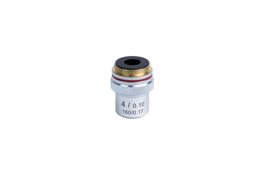
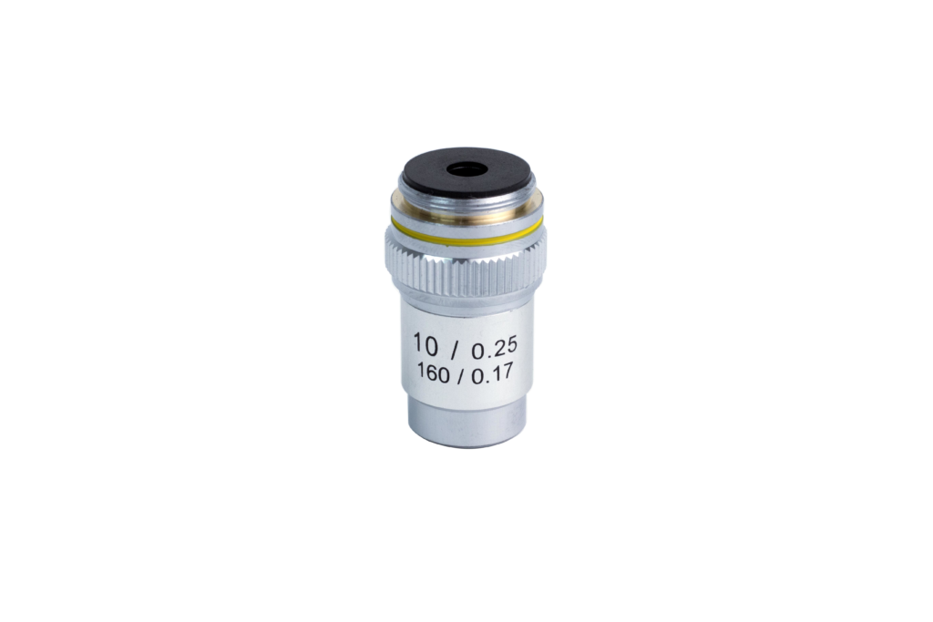
For newbies and people not using microscopes frequently, the lowest magnification setting has the largest field of view, so it is the easiest to locate your microscopic sample and center on it. It’s also the easiest to focus, so you can start there, then change the objective to the next higher, refocus, re-center, and repeat until the desired magnification setting is achieved. This would increase your confidence in using the microscope, and avoid any frustration you might have for not locating or focusing the sample properly.
If you are a microscope veteran and can immediately focus and scan on higher magnification values, go for it. There is no hard and fast rule that says you HAVE TO start at 4x or even lower. However, it’s just easy to most novices to star that way, so we recommend doing so.
c
WHAT MAGNIFICATION DOES MY MICROSCOPE NEED TO BE ABLE TO SEE CELLS?
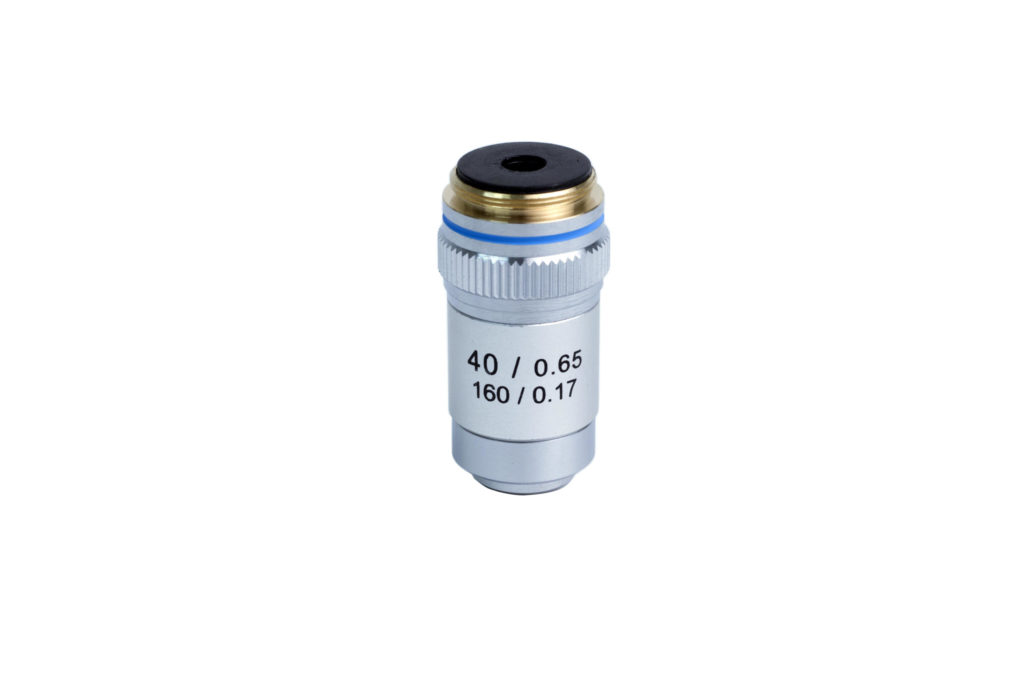
Typically, not only will you need a compound microscope (a microscope with an illuminator at the base of the microscope that shines up through the slide, into the lens, and creates an image in the eyepiece), but you will need one that will operate to a minimum magnification of 400x (or a 10x eyepiece with a 40x objective).
WHAT MAGNIFICATION DOES MY MICROSCOPE NEED TO BE ABLE TO SEE BACTERIA?
Bacteria are also cells (typically single cell organisms). Again, a compound microscope with a minimum magnification of 400x is the most common combination you will need to see the ubiquitous, mostly free-living organisms: bacteria.
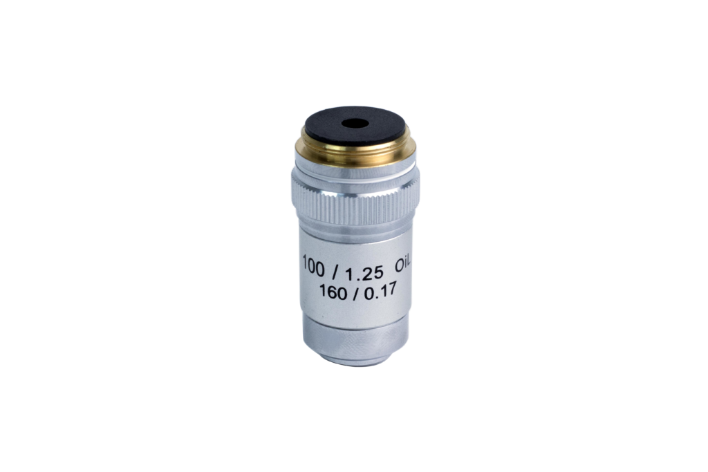
The higher magnification, the more detail you will see (up to 1000x total magnification). Beyond 1000x, you will enlarge the image but you won’t add more detail (more magnification, no additional resolution).
WHAT IS A MECHANICAL STAGE?
A microscope stage on a compound microscope has to move vertically up and down in order to achieve focus, so it is too simplistic to say a mechanical stage is one that moves. There are 2 types of microscope stage on a compound microscopes: stage clips and mechanical stage. While the stage clips are used to hold the microscope slide in place and can’t be moved, the mechanical stage can move in the X and Y dimensions via 2 knobs in order to allow a user to scan through a sample, or easily center it in the filed of view without using by hand.
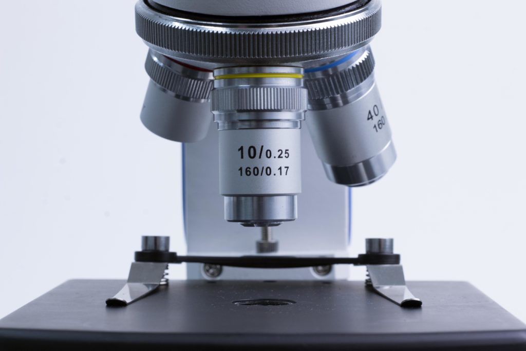
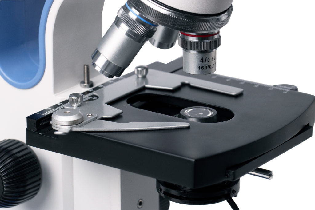
Is the mechanical stage better than the stage clips? Absolutely! Using stage clips can cause frustration as you have to move the slide up and down, left to right by your hand(s) to locate the sample and focus. Touching a slide by hand is both cumbersome, clumsy, and can leave oils that disturb the image you are trying to view. Hence, a mechanical stage is highly sought after in a quality microscope.
WHAT IS IRIS CONDENSER? & WHAT DOES IT DO?
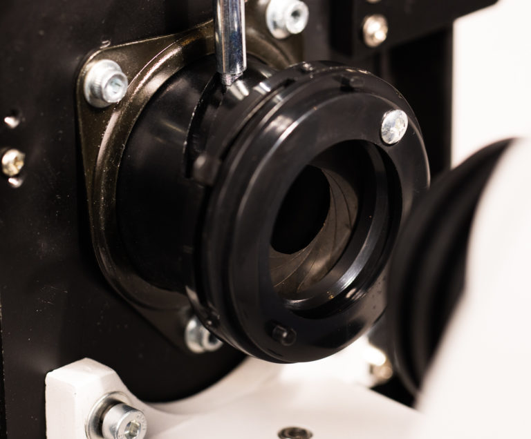
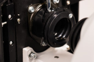
The condenser is used to collect and focus the light from the illuminator onto the sample. It is located under the stage often in conjunction with an Iris diaphragm which controls the amount of light reaching the sample. The Iris in the condenser allows you to see the depth of field when focusing on a slide, most subjects will have depth of field unseeable with the naked eyes, under high magnification it can be seen.
By closing the Iris down, it allows you to focus on the closest and the furthest part of the sample. When the Irises is closed, you require brighter illumination.
WHAT IS BETTER, LED OR HALOGEN?
There isn’t a solid answer here as it’s mostly personal preference. In general, it depends on you application (like most things microscopy related).
Pros of Halogen bulbs:
- are common in microscopy
- cheap and replaceable bulbs
- provide close to natural sunlight (even more so with a daylight blue filter)
Cons of Halogen bulbs:
- Emit a great deal of heat when used, which can limit how long you can view a liquid or heat sensitive sample before it becomes dried up and dies
- the bulbs do burn out so they have to be replaced quite frequently if you use the microscope for a long time or in a frequent basis.
Pros of LED:
- are used frequently these days due to its extremely long lasting nature
- low power draw
- lack of heat emission
Cons of LED:
- they are soldered onto a board of their own in order to function
- they are often a bright white light that can be slightly discolour samples instead of the natural sunlight yellow of a halogen.
WHAT IS A DIOPTER ADJUSTMENT ON A MICROSCOPE USED FOR?
Diopter Adjustment on the eyepiece. Interpupillary adjustment (width between eyes) shown by the arrow at the bottom.


If you have a fixed eyepiece and 1 diopter, focus the microscope, then look into it with the eye that is on the fixed ocular tube side (typically right). From there, look in the opposite eye, and adjust diopter until the adjustable eye becomes focused. That’s all there is to it!
If you have 2 adjustable diopters, follow the same process. Set 1 dead center in the travel range, and adjust the other until both are clear. you may need to go back and forth adjusting, which is fine. Just take your time to be sure that it is correct for you to avoid eye strain and headaches!
You can also use the diopter adjustment to help collimate the image-basically, making the 2 circles of the images from each eye appear to be one. If each eye seems to see an individual image, give this an adjustment.
SHOULD I GET A MONOCULAR/ BINOCULAR OR A TRINOCULAR MICROSCOPE?
“Ocular” refers to the eye, and thus, the tube in which an eyepiece or camera goes into to allow a user to view into the microscope. The prefixes determine the number of ocular tubes on a microscope -“mono-” meaning one, “bi-” meaning two, and “tri-” meaning three.
If the microscope is primarily to be used by students from ages K-12, a monocular microscope would be the most ideal option. Many binocular microscopes are a bit too large for younger students to use. Even though the interpupillary distance can be adjusted, often it cannot be adjusted small enough to fit their smaller widths between their eyes. Equally important, you can save up over $200 ea by purchasing a monocular microscope over a binocular microscope as well.
References
MicroscopeGenius, 2015, “15 Answers for Common Microscope Newbie Questions”, weblog post, 22 October, viewed 20 June 2021, <https://microscopegenius.com/15-answers-for-common-microscope-newbie-questions/>
Levenhuk, n.d, “Answers to frequently asked questions about microscopes”, weblog, view 10 May 2021, <https://www.levenhuk.com/reviews/answers-to-frequently-asked-questions-about-microscopes/>
